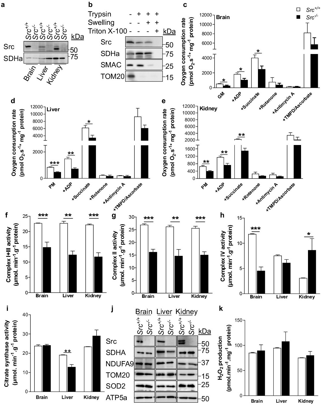
Fig. 1. Deletion of Src impairs mitochondrial metabolism in brain, liver and kidney. (a) Representative immunoblots (n=4) of Src and SDHa (as loading control) in brain, liver and kidney mitochondria isolated from Src+/+ and Src-/- mice (fed ad libitum). See Fig. S1a for quantification. (b) Representative immunoblots (n=3) of Src, the inner mitochondrial membrane protein SDHa, the mitochondrial intermembrane space protein SMAC and the outer membrane mitochondrial protein TOM20 in liver mitochondria treated as indicated, showing that Src is mostly located inside mitochondria. (c-e) Oxygen consumption rates of brain (c), liver (d) and kidney (e) mitochondria isolated from Src+/+ and Src-/- mice (fed ad libitum) in the presence of different substrates and inhibitors, as indicated (n=4). GM: Glutamate/Malate, PM: Pyruvate/Malate. (f-i) Enzymatic activities of complexes I+III (f), II (g) and IV (h), and citrate synthase (i) in brain, liver and kidney mitochondria isolated from Src+/+ and Src-/- mice fed ad libitum. (j) Representative immunoblots (n=4) of Src, SDHa, NDUFA9, TOM20, SOD2 and ATP5a in brain, liver and kidney mitochondria isolated from Src+/+ and Src-/- mice fed ad libitum. See Fig. S1b for quantification. (k) H2O2 production by brain, liver and kidney mitochondria isolated from Src+/+ and Src-/- mice fed ad libitum (n=4). Data are presented as mean ± s.e.m. *p<0.05, ** p<0.01, ***p<0.001 determined by Student's T test.